Human-Centered Design Principles
Creating a user-centered and efficient user interface (GUI) for this clinical application that generates real-time 3D maps of the heart is crucial for accurate diagnosis and treatment.
Conducted usability testing with healthcare professionals to gather feedback and refine the UI based on their real-world usage and requirements.
Discovered user-needs and requirements
• Make the 3D heart mapping the central element of the UI.
• Use high-quality graphics and allow users to rotate, zoom, and pan to examine the heart from different angles.
• Display real-time updates of the heart's condition, including vital signs, electrical activity, and any anomalies.
• Use color coding and visual cues to highlight abnormalities.
• Allow user to customise their preferred views of the 3D heart model. They should be able to save presets for different perspectives and measurements.
• Allow users to add editable annotations and markers directly on the 3D model to highlight specific areas of interest.
• Integrate patient data, ECG readings, and other relevant metrics.
• Include navigation tools like a scrubber feature to help users to pinpoint specific moments during the procedure.
• Provide the option to open multiple windows for side-by-side comparisons of different views of the model.
• Provide users with guidance to help them quickly become familiar with the application's features.
Icon family design
Heart conditions show up as irregularities in the organ’s electrical routine. Heart surgeons and physicians sometimes check in on that routine by using catheters with electrodes at their tips. They wind up a vein or artery in the patient’s legs or arms and into their heart chambers where they record electrical signals thundering through the muscle.
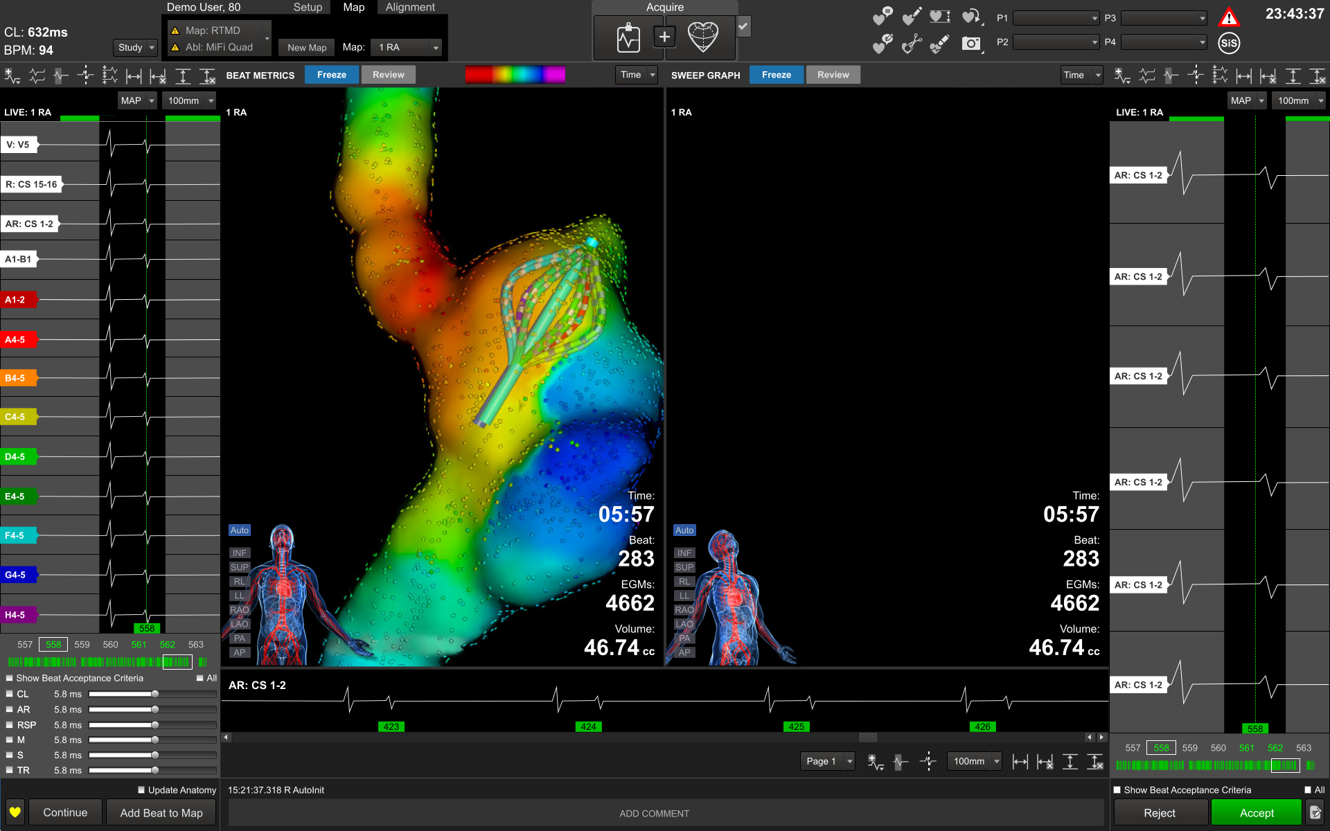
The system translates mere pings into pixels to give doctors a clearer picture of the heart’s architecture and electrical activity–in 3-D–as it pumps.